Tuesday, March 30, 2010

Osteochondral Fracture


The procedure that took place this morning was the removal of an osteochondral defect of the left lateral femoral condyle; and it was a rather large piece! The patient, 56 male, initially reported a feeling of "locking," and "intense pain" along the medial and lateral joint line, also with stiffness deep from within the knee joint, and some "instability." Upon evaluation of radiographs, the Doctor felt there may be a presence of loose bodies, commonly referred to as 'joint mice,' or technically termed osteochondral defects.

However, while the knee was being scoped, the Doctor noticed a particularly large piece on the monitor. It took some time for him to grab a hold of it, even at one point, losing sight of it and having to reconfigure himself and enter from a different angle. Once he got a decent view of the object, he realized how large of a piece it was. He struggled for next few minutes to finally get a hold of the piece and remove it.

Once removed, he looked around the condyles and any other structures to get an idea where such a large piece may have come from. There was no definitive answer. At this point, they decided to have the knee x-rayed and maybe that could help them discover where it came from, also, if there were any other pieces around they did not get.

There were a few moments that took me by surprise; a "whoa" moment, if you will. There was a point where the surgical table was too high, and he asked for it to be lowered, and apparently the table was being lowered, but the technician didn't know how to stop it, so the patient was being lowered while there were surgical tools still inside him. It may not have been much of a deal, but I was certainly taken away at the time. The other thing that happened, which gave me more insight into the risks with surgery, was the patient was bleeding more than normal, and the doctor requested the tourniquet's pressure be increased. Again, one of the tech's was unsure on how to do this, or maybe just wasn't familiar with the equipment or could not locate the proper tool to, etc., I am not sure, but the Doctor sure was upset. And I am guessing, the last thing you want, as a patient, is to have an angry Doctor performing your surgery.

Wednesday, March 24, 2010

TheraLase Presentation Summary


I thoroughly enjoyed the presentation given this morning on Thera Lase, which is a new product of low level laser therapy, typically used for chronic pain, wounds, and tissue recovery. The presentation was very well done, professionally given, easy to follow, and did an excellent job of explaining its use.

One of the most surprising claims the presenter made that really caught my attention was its effects on fungal infections similar to eczema, or like tinea versicolor, which is a skin condition I have had for 13 years. This was surprising to me at the time, and although I did not follow up with a question, I did my own research on the companies' website.

Upon navigating their website, there is a section on Anti-Aging Treatments, which is the closest category I could find to skin conditions. There, they talk about reducing lines and wrinkles, crows feet, blemishes, acne, scars, etc., no mention of treatment of fungal conditions. All the text says are relative to surface layer treatments and conditions whereas tinea versicolor is treated more on an internal level.

After a little more searching, I steered over to the clinical practitioner section of the website, where I discovered a little more of the information I was seeking. Although for most part, it says the same thing as the patient section, however, it gives a little more scientific background mentioning that its use increases local vascularity and capillary formation, which in turn, brings more oxygen and nutrients to the area, in order to stimulate collagen growth. There is also a list of treatable conditions that include; herpes simplex, psoriasis, rosacea, keloids, stretch marks, and others I mentioned above; but I noticed how none of those conditions are caused by a fungus. So, it is still unclear what they meant as to what skin conditions are treated with the laser. The only other explanation would be if I mistakenly heard one word for another.

Thursday, March 18, 2010

Total Hip Replacement

The procedure I observed this morning was a right total hip replacement, in which the patient was a 79 year old woman, whom had her left hip replaced five years prior. The surgery was scheduled to begin at 7:00 AM, however, the first incisions were not made until 8:45AM. Other than the Doc being slightly late, there was a staggering amount of preparation that went into this surgery, which may have contributed to the delay. The technicians scrubbed the patients' entire leg for a good ten minutes before wrapping it in the necessary materials to prepare for the surgery.

The patient was unquestionably a larger woman which proved to be no obstacle for the Doctor, as he navigated his way through skin, adipose tissue, and muscle to locate the greater trochanter, and eventually the femoral head which was subsequently sawed off. Unfortunately, the pictures I took during the procedure were all on my phone, which was recently destroyed, otherwise I would show a few great shots of the incisions and the sawed-off femoral head.

The procedure itself was pretty straight forward. There wasn't too many interesting things that took place while in the surgery room. Every now and then, the Physician Assistant would toss a quiz question my way, and since there was another high school student in attendance, the Physician Assistant allowed me to answer some questions, however, he shot most of the questions to the high school student, because "he hadn't already had anatomy," and I have, so he expected me to know all the answers to his questions.

Other interesting things that happened during the surgery was one of the technicians apparently became uneasy during the surgery and needed to be helped out of the room. She did not pass out, per se, but from the looks of it, she was well on her way to taking a dive into the operating rooms' floor.

There were a few moments during the middle of the procedure where the Physician Assistant had to shift and adjust the patients' leg positing, which was 1) so the Doctor could get a better view, and 2) so they can observe the way the newly added hardware would work with the patients' existing bony structures, in which they simply simulated all the motions of the hip. There was one time when the Physician Assistant joked, "I will give anyone in here $20 if they could position their leg like this," and I saw just exactly how he was holding the leg, and there is no one alive who could do that. The leg was placed into hip adduction, internal rotation, knee flexion, and and horizontally adducted to where the involved foot could reach the opposite side (right ASIS) and then bend upwards. It was certainly a freakish sight to see and very unnatural, of course.

Needless to say, the environment in their operating room was very relaxed and inviting. There were several times when the Doctor and the Physician Assistant allowed us to get closer looks and really, truly allowed us the opportunity to learn.

Saturday, March 13, 2010

Familiar face?


Came across this article on Huffpost and couldn't help but recognize the face in the background -- is that you Father Privett, President of my alma mater?


Sunday, March 7, 2010

An amazing tale of heroism

...or just another day in the life of an Athletic Trainer!

This afternoon while attending Ben's usual Sunday soccer game (he participates in a Mexican League), we witnessed a pretty nasty injury to one of the opposing players. In the late second half, the score was 3-2 so both teams are battling; one team to tie the game, the other to win the game. Lauren P. (also another emergency responder) and I are sitting in upper part of the stands when these two teammates both go for the ball. I'm presuming they didn't see one another. One teammate goes to kick the ball, but instead, kicks his own guy along the side of his lower leg. The teammate then proceeds to fly about 4ft. in the air, inverts head-over-heel, and lands on his back. However, while in the air, the kids' foot points outwards, as if it was broken in half -- just dangling there, prior to him slamming back into the ground. Lauren and I both have an "Oh S__t!" moment. Well, her's was more like a "Oh my!," but still we both had sudden reactions from the sight of the grossly deformed foot.

At that moment I run onto the field to check on the athlete. He is visibly hurt and in pain, yet calm. Some of the players begin to surround the scene. I survey the athlete and his foot does not appear to be in any unnatural position. In fact, it is pointed straight and not out to the side, like it was when upon initial contact. The player is on his side, as pictured below, and reaches down to grab for his leg. Upon closer inspection, there appears to be a bulge along the lateral malleous, or outside ankle, apparently the site of injury.

There were over 300+ Spanish speaking individuals (a language which I do not speak fluently) so I motioned to one of the persons standing by for his shirt, or jacket, to cover up the foot, for fear that the player would see it and go into shock. It was just a precautionary measure, but luckily, the player was very calm. He squirmed and groaned occasionally, but overall, he was very calm.

By now, Ben and I are both at the scene, some 1.5 minutes into the ordeal, and the the players' cousin, is dialing 911, but no one (out of the 300+ people out there) knew exactly where we were!


Meanwhile, Ben begins to palpate the players ankle and lower leg feeling for deformities. I report to him what I saw, and he continues his inspection. By now, everything has stopped (on both fields) and everyone has gathered around, so I step back and try to restore control, making space for Ben to work and creating a path for the ambulance. The cousin on the phone finally communicates to the authorities that we are at a park off the loop, that when I say "Near the expo center," which would hopefully give them a better idea of our location.

Ben is busy checking for breaks in continuity, checking pulses, and making sure everything is intact, while we wait for the ambulance to arrive. He is also asking questions about relevant sounds and sensation, all through the cousin who is translating every word. At this point, the only thing we could do is manage the scene, keep the athlete as comfortable as possible (placing a rolled jacket underneath his head), monitor his consciousness making sure he's not slipping into shock (he said he didn't want the jacket we offered to keep him warm), and wait for the Emergency Personnel to arrive.


It was at this point where I pulled out my phone and began to take pictures, and Ben instructs one of the persons standing by to flag down the ambulance once it arrives. They got there quickly in under 12-13 minutes.


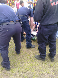
Emergency personnel working to remove the shoe.


Ben (blue white stripes) assisting with the care of the athlete. A closer look reveals the position of the splinted foot, wrapped in what appears to be a rigid "L" shape splint that covers the foot posteriorly, with a soft cushion material protecting it anteriorly and laterally, which had the appearance of an Ace bandage.


Once the players shoe was removed, he was lifted and boarded onto the stretcher and placed into the ambulance. And 5 minutes later, the game resumed. Ben's team ended up losing 3-2.


Video of Ben's inspection and communicating with the cousin, right before the ambulance arrives.

My impressions of what happened, and over reflection with Ben, is that the guy suffered a left leg distal fibular fracture, evidenced by the 90 degree angulation his foot presented with while he was in the air, but possibly upon impact with the ground, his foot shifted to its normal position, although by normal, I mean it was no longer pointed outwards. During Ben's inspection he noticed some discontinuity over the lower fibular shaft, and noticeable deformity, but it was a closed-fracture, or there was no skin breakage.